At-Home CRP Tests: What They Measure and How to Read Them
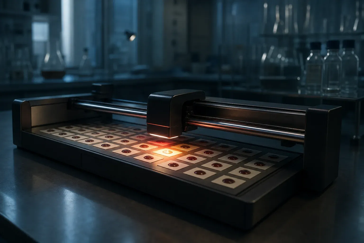
Yes. You can measure C-reactive protein at home three ways: a finger-prick sample you collect and mail to a lab, a dried blood spot card, or a lateral-flow or point-of-care cartridge that reads out in ten minutes. The one thing that matters for choosing among them is the assay’s lower limit. Standard CRP assays report down to about 3–5 mg/L and are built for infection and flare monitoring. High-sensitivity CRP (hs-CRP) assays report down to roughly 0.1–0.2 mg/L and are the ones used for cardiovascular risk stratification, where the entire question lives between 0.5 and 3 mg/L. If a kit says “CRP” without “hs-” and quotes a range starting at <5 mg/L, it cannot answer the question most technical readers are asking.
There is no chemistry difference worth caring about. Both are immunoturbidimetric or immunonephelometric assays against the same pentameric protein, usually latex-enhanced. hs-CRP just means the calibration curve and the detector were built for the low decade. Some labs run one assay and report it over the full range, which is the ideal case.
Which format to use, and why we prefer mail-in serum or plasma
If you want a number you can put in a time series and compare to a paper, order a mail-in kit that ships your sample to a CLIA lab running a hs-CRP immunoassay, and ask for the numeric result plus the assay name. You get a continuous value with about 3–5% analytical CV, a documented reference interval, and a result that is comparable to any hospital draw you have ever had.
The alternatives, ranked by how much I trust the number:
Dried blood spot. You bleed onto filter paper, let it dry, mail the card at ambient temperature. This is the format used in field epidemiology because it survives shipping without cold chain. CRP from DBS correlates well with serum, but the absolute values need a conversion equation and the hematocrit of your spot affects the plasma fraction you are eluting. A field validation of DBS CRP in household air pollution studies found the method usable for population comparisons and reported the correlation and the practical handling failure modes, including spot volume and humidity exposure.1 For self-tracking, DBS is fine as long as you keep the same vendor and the same assay, so your bias is constant.
Point-of-care cartridge. Whole-blood immunoassay, result in 3–10 minutes, often semi-quantitative (bands like <10, 10–40, >40 mg/L) or quantitative with a wide CV. These were designed for a specific clinical job: deciding at the bedside whether a patient with a respiratory complaint needs antibiotics or hospital referral. The literature on POC CRP as a primary-care triage tool is exactly that use case.2 Semi-quantitative bands are useless for hs-CRP work. A band of “<10” contains the entire interesting range.
Silicon photonics home platforms. Newer at-home analyzers run multiplexed immunoassays on a photonic chip from a finger-prick sample, with CRP typically in the panel. A published characterization of one such platform reports measurement of blood biomarkers at home with correlation against reference laboratory methods.3 These are worth watching because they give you frequent sampling without shipping. Check the reported lower limit of quantitation for CRP specifically before you assume it is hs-grade.
The single biggest interpretation error: sampling once
CRP has enormous within-person variability. It is an acute-phase reactant with a plasma half-life of about 19 hours and a hepatic production response that can climb more than a thousand-fold from baseline. A single 2.8 mg/L result tells you almost nothing about your set point. A dental infection, a sprained ankle, a viral cold three days ago, a poorly slept week, or a hard eccentric training session will all move it.
What we do: three measurements, at least two weeks apart, none within 10 days of a known infection or an unusual training load, and take the median. Then repeat quarterly. The signal you want is the drift in your median across years, not the individual points. If two of three baseline draws are above 3 mg/L with no explanation, that is a conversation with a clinician, not something to interpret from a blog post.
Practical logging schema, because you will want this structured from day one:
date, time_local, assay ("hs-CRP" | "CRP"), vendor, matrix ("serum"|"plasma"|"DBS"|"whole_blood"),
value_mg_L, lloq_mg_L, fasting_hours, days_since_symptoms, hard_exercise_48h (bool),
sleep_hours_prior, notes
The lloq_mg_L field matters more than people expect. A result reported as “<1.0” from one vendor and “0.4” from another are not different biology.
What moves CRP, in rough order of effect size
Acute infection dominates everything. Bacterial infection commonly drives CRP into the tens or hundreds of mg/L. In experimental human endotoxemia and in severe sepsis, the acute-phase and endothelial damage markers rise together on a timescale of hours, which is why CRP alone is a lagging and non-specific sepsis indicator.4 CRP does not peak until 36–50 hours after the insult, so in the first day of a serious illness it can be misleadingly normal. Neonatal sepsis work has looked for markers with better early sensitivity than CRP for the same reason.5
Adiposity, chronic low-grade inflammation, and fitness set the baseline. Structured exercise programs measurably lower inflammatory markers: a randomized multicomponent exercise trial in older community-dwelling adults reported reductions in inflammatory biomarkers alongside improvements in physical performance.6 Expect months, not weeks.
Air pollution and inhaled particulate exposure. Studies in COPD patients have examined the association between air pollution exposure and systemic inflammation and tissue repair markers.7 If you live somewhere with wildfire smoke seasons, annotate your log.
Cardiovascular relevance. CRP is one of several inflammatory markers studied in coronary artery disease, and the reviews are candid that its predictive value is modest and additive rather than decisive.8 It is a covariate, not a verdict.
Respiratory viruses. CRP rises with viral infection too, just less dramatically than with bacteria, and other mediators separate the causes better. Serum IP-10 has been studied as a marker of rhinovirus infection at COPD exacerbation, which is the kind of specificity CRP does not have.9
Conventional interpretive bands
For hs-CRP measured at baseline health, the widely used tertiles are <1.0 mg/L (low), 1.0–3.0 (average), >3.0 (high). Above 10 mg/L, stop treating the number as a risk marker: that is an acute-phase response, and the right move is to find the cause or wait two weeks and remeasure. Values in the hundreds belong to acute care, not self-tracking.
CRP is also a poor standalone discriminator in the settings where it is measured most. In periprosthetic joint infection, for example, hematologic ratios have been evaluated precisely because CRP and ESR alone leave too much diagnostic uncertainty.10 Pair CRP with a CBC with differential, ferritin, and albumin if you want context. All four move together in inflammation, and ferritin plus albumin help you tell “CRP is up because of an acute-phase response” from assay noise.
If you have a CRP above 10 mg/L with fever, confusion, rapid breathing, or low blood pressure, that is an emergency and you should be seen immediately, not remeasuring at home.
Questions people also ask
Can I test my CRP at home? Yes, via mail-in finger-prick, dried blood spot cards, or a point-of-care cartridge. Insist on a quantitative hs-CRP result with a stated lower limit of quantitation if your interest is cardiovascular risk rather than infection monitoring.
Can CRP be high without infection? Routinely. Obesity, recent intense exercise, smoking, poor sleep, autoimmune activity, trauma, surgery, periodontal disease, and particulate air pollution exposure all raise it. CRP tells you an acute-phase response is happening, not what triggered it.
Can anxiety cause high CRP levels? Chronic psychological stress is associated with modestly higher inflammatory markers in population studies, generally a fraction of a mg/L in effect size. It does not explain a result in the tens.
How serious is a CRP of 300? That is a severe acute-phase response, typically bacterial infection, major trauma, or surgery, and it is a hospital-level finding. Get evaluated in person.
How much CRP indicates sepsis? There is no threshold that establishes sepsis. CRP lags the insult by a day or more and rises in many non-infectious states, so sepsis is defined by organ dysfunction and clinical criteria with cultures and lactate, not by a CRP cutoff.4
What autoimmune disease causes high CRP? Rheumatoid arthritis, the spondyloarthropathies, and vasculitides typically raise CRP markedly during flares. Systemic lupus is the notable exception, where disease activity often occurs with a normal or only mildly raised CRP and a high ESR, which is one reason the discordance itself is informative to a rheumatologist.
Oak builds longitudinal molecular profiles of individuals: whole-genome sequencing, RNA sequencing, proteomics, blood biomarkers, and continuous glucose data, integrated into one model of you. Build your profile.
Footnotes
-
Bonnie N. Young, Jennifer L. Peel, Tracy L. Nelson, et al. C‐reactive protein from dried blood spots: Application to household air pollution field studies. Indoor Air, 2019. https://doi.org/10.1111/ina.12603 ↩
-
T Scott Isbell. Home or Hospital? Point-of-Care CRP as a Triage Biomarker in the Primary Care Setting. Clinical Chemistry, 2017. https://doi.org/10.1373/clinchem.2016.269159 ↩
-
Kyle J. Preston, Guojun Chen, Cole A. Chapman, et al. At-home measurement of blood biomarkers on the SiPhox Home silicon photonics platform. Frontiers in Biological Detection: From Nanosensors to Systems XVI, 2024. https://doi.org/10.1117/12.3001255 ↩
-
Sisse R. Ostrowski, Ronan M.G. Berg, Nis A. Windeløv, et al. Coagulopathy, catecholamines, and biomarkers of endothelial damage in experimental human endotoxemia and in patients with severe sepsis: A prospective study. Journal of Critical Care, 2013. https://doi.org/10.1016/j.jcrc.2013.04.010 ↩ ↩2
-
Tai-Wei Wu, Meredith Tabangin, Ryosuke Kusano, et al. The Utility of Serum Hepcidin as a Biomarker for Late-Onset Neonatal Sepsis. The Journal of Pediatrics, 2013. https://doi.org/10.1016/j.jpeds.2012.06.010 ↩
-
Uratcha Sadjapong, Supachai Yodkeeree, Somporn Sungkarat, et al. Multicomponent Exercise Program Reduces Frailty and Inflammatory Biomarkers and Improves Physical Performance in Community-Dwelling Older Adults: A Randomized Controlled Trial. International Journal of Environmental Research and Public Health, 2020. https://doi.org/10.3390/ijerph17113760 ↩
-
Payam Dadvand*, Mark J Nieuwenhuijsen, Àlvar Agustí, et al. Air Pollution and Biomarkers of Systemic Inflammation and Tissue Repair in COPD Patients. ISEE Conference Abstracts, 2014. https://doi.org/10.1289/isee.2014.p3-683 ↩
-
Epaminondas Zakynthinos, Nikolitsa Pappa. Inflammatory biomarkers in coronary artery disease. Journal of Cardiology, 2009. https://doi.org/10.1016/j.jjcc.2008.12.007 ↩
-
Jennifer K. Quint, Gavin C. Donaldson, James J.P. Goldring, et al. Serum IP-10 as a Biomarker of Human Rhinovirus Infection at Exacerbation of COPD. Chest, 2010. https://doi.org/10.1378/chest.09-1541 ↩
-
Venkatsaiakhil Tirumala, Christian Klemt, Liang Xiong, et al. Diagnostic Utility of Platelet Count/Lymphocyte Count Ratio and Platelet Count/Mean Platelet Volume Ratio in Periprosthetic Joint Infection Following Total Knee Arthroplasty. The Journal of Arthroplasty, 2021. https://doi.org/10.1016/j.arth.2020.07.038 ↩